Editor-in-Chief : V.K. Rastogi

ASIAN JOURNAL OF PHYSICS
An International Peer Reviewed Research Journal
Frequency : Monthly,
ISSN : 0971 – 3093
Editor-In-Chief (Hon.) :
Dr. V.K. Rastogi
e-mail:[email protected]
[email protected]
| AJP | ISSN : 0971 – 3093 Vol 26, No 11-12, November-December, 2017 |
Asian
Journal of Physics
|
Asian Journal of Physics |
Vol. 26 No 11 & 12 (2017) 325-335 |
The anti-HIV Nucleoside analogue d4T (Stavudine): Solid state simulation by
DFT methods of the FT-IR and FT-Raman spectra
M Alcolea Palafoxa,b, D Kattana,b, and A Nils Kristiana
aNofima AS – the Norwegian Institute of Food, Fisheries and Aquaculture Research, Osloveien 1, 1430 Ås, Norway
bDepartamento de Química-Física I, Facultad de Ciencias Químicas,Universidad Complutense, Madrid-28040, Spain
The theoretical and experimental vibrational study of the anti-HIV d4T (stavudine or Zerit or 3′-deoxy-2,3´-didehydro thymidine) Nucleoside Analogue was carried out. The calculated spectra were scaled by using the linear scaling equation procedure (LSE). The d4T monomer and dimers were simulated by using DFT methods. The IR spectrum was recorded in the solid state in the region 400-4000 cm-1 and the Raman spectrum was recorded in the region 0-3500 cm-1. The vibrational bands were analyzed and assigned to different normal modes of vibration by comparison with the scaled values of the different dimer forms. Thus, through this comparison, we were able to confirm that the solid state sample corresponds to dimer V. © Anita Publications. All rights reserved.
Keywords: d4T, Stavudine, anti-HIV, IR spectrum, Raman spectrum, DFT
References
1. Taisheng L, Fuping G, Yijia L, Chengda Z, Yang H, Wei L, Yun H, Hongzhou L, Jing X, Aiqiong H, Yanling L, Xiaoping T, Hui W, Tong Z, Guiju G, Junkang L, Xiaoying Z, Xinhua W, Yongtao S, Jinsong B, Ling L, Huanling W, Chinese Med J, 127(2014)59-65.
2. Li T, Dai Y, Kuang J, Jiang J, Han Y, Qiu Z, Jing Xie, Zuo L, Li Y, PLoSONE 3, e3918 (2008); doi.org/10.1371
3. van Oosterhout J J, Mallewa J, Kaunda S, Chagoma N, Njalale Y, E. Kampira E, Mukaka M, Heyderman R S, PLoSONE 7, e42029(2012); doi.org/10.1371 /journal.pone.0042029
4. Phanuphak N, Ananworanich J, Teeratakulpisarn N, Jadwattanakul T, Kerr S J, Chomchey N, Hongchookiat P, Mathajittiphun P, Pinyakorn S, Rungrojrat P, Praihirunyakit P, Gerschenson M, Phanuphak P, Valcour V, Kim J H, Cecilia S, Antivir Ther, 17(2012)1521-1531.
5. Palafox M A, Iza N, J Molec Struct, 1028(2012)181-195.
6. Palafox M A, Talaya J, J Phys Chem B, 114(2010)15199-15211.
7. Zhao Y, Truhlar D G, J Chem Phys, 125(2006)194101; doi:10.1063/1.2370993.
8. Frisch M J, Trucks G W, Schlegel H B, Scuseria G E, Robb M A, Cheeseman J R, Scalmani G, Barone V, Mennucci B, Petersson G A, Nakatsuji H, Caricato M, Li X, Hratchian H P, Izmaylov A F, Bloino J, Zheng G, Sonnenberg J L, Hada M, Ehara M, Toyota K, Fukuda R, Hasegawa J, Ishida M, Nakajima T, Honda Y, Kitao O, Nakai H, , Vreven T, Montgomery J A(Jr), Peralta J E, Ogliaro F, Bearpark M, Heyd J J, Brothers E, Kudin K N, Staroverov V N, Kobayashi R, Normand J, Raghavachari K, Rendell A, Burant J C, Iyengar S S, Tomasi J, Cossi M, Rega N, Millam J M, Klene M, Knox J E, Cross J B, Bakken V, Adamo C, Jaramillo J, Gomperts R, Stratmann R E, Yazyev O, Austin A J, Cammi R, Pomelli C, Ochterski J W, Martin R L, Morokuma K, Zakrzewski V G, Voth G A, Salvador P, Dannenberg J J, Dapprich S, Daniels A D, Farkas Ö, Foresman J B, Ortiz J V, Cioslowski J, Fox D J, Gaussian 09, Revision D.01, Inc., Wallingford CT, 2009.
9. Palafox M A, Rastogi R, Anupama, Alam M J, Bhat D, Rastogi V K, Asian J Phys, 25(2016)189-219.
10. El-Sayed A A, Molina A M, Álvarez-Ros M C, Palafox M A, J Biomol.Struct & Dyn, 33(2015)723-748.
11. Palafox M A, Chem Informatics, 1(2015)1-13.
12. Palafox M A, Posada-Moreno P, Villarino-Marín A L, Martinez-Rincon C, Ortuño-Soriano I, Zaragoza-García I, J Computer-Aided Molec Design, 25(2011)145-161.
13. Palafox M A, Iza N, Struct Chem, 24(2013)967-980.
14. Yurenko Y P, Zhurakivsky R O, Ghomi M, Samijlenko S P, Hovorun D M, J Phys Chem B, 111(2007)6263-6271.
15. Palafox M A, Rastogi V K, Spectrochim Acta A, 58 (2002)411-440.
16. Choi Y, George C, Comin M J (Jr). Barchi J J, Kim H S, Jacobson K A, Balzarini J, Mitsuya H, Boyer P L, Hughes S H, MarquezV E, J Med Chem, 46(2003)3292-3299.
17. (a) Palafox M A, Int J Quantum Chem, 77(2000)661-684.
(b) Palafox M A, Rastogi V K, in Perspectives in Modern Optics and Optical Instrumentation, (eds), Jobi J, Sharma A, Rastogi V K, (Anita Publications, FF-43, Mangal Bazar, Laxminagar, Delhi), 2002, pp 91-98
(c) Palafox M A, Nunez J L, Gill M, Rastogi V K, in Perspectives in Engg Optics,, (eds) Singh K, Rastogi V K, (Anita Publications, FF-43, Mangal Bazar, Laxminagar, Delhi), 2003, pp 356-391.
(d) Palafox M A, Nunez J L, Gill M, Rastogi V K, Mittal L, Sharma R, Int J Quantum Chem, 103(2005)394-421.
(e) Palafox M A, Asian Chem Letts, 7(1998)785-816.
18. Palafox M A, Iza N, Gil M, J Mol Struct (Theochem), 585(2002)69-92.
19. Rastogi V K, Singhal S, Palafox M A, Rao G R, Ind J Phys, 84 (2010)151-165.
20. Carpenter J E, Weinhold F, J Molec Struct, (Theochem) 169(1988)41-62.
21. Alvarez-Ros M C, Palafox M A, Pharmaceuticals, 7(2014)695-722.
22. Palafox M A, Struct.Chem, 25(2014)53-69.
23. Palafox M A, J Biomol.Struct & Dyn, 32(2014)831-851.
24. Alvarez-Ros M C, Palafox M A, J Molec Struct, 1047(2013)358-371.
25. Gauss View 5.1 Gaussian Inc., Wallingford CT, (2009).
26. Saenger W, Principles of Nucleic Acid Structure, (Springer Verlag Publishers: New York),1984, pp 556.
27. Mirmehrabi M, Rohani S, Jennings M C, Acta Cryst, C61(2005)o695-o698; doi.org/10.1107/S01082701050345918
28. Gurskaya G V, Tsapkina E N, Abstracts of the twelfth European Crystallographic Meeting, Moscow, vol. 2, p 380(1989).
29. Tsapkina E N, Dissertation for Doctorate I Chemical Sciences, Institute of Molecular Biology, Moscow, (1989).
30. Harte W E (Jr), Starret J E (Jr), Martin J C, Mansuri M M, Biochem Biophys Res Comm, 175(1991)298-304.
31. Gurskaya G V, Bochkarev A V, Zhdanov A S, Dyatkina N B, Kraevskii A A, Mol Biol., 25(2),401 (1991).
32. Rastogi V K, Singh C, Jain V, Palafox M A, J Raman Spectrosc, 31(2000)1005-1012.
33. C J Cramer, Essentials of Comput. Chem.;( John Wiley & Sons: Chinchester, England), 265 (2002).
34. Palafox M A, Iza N, Gil M, J Molec Struct (Theochem), 585(2002)69-92.
35. Colarusso P, Zhang K, Guo B, Bernath P F, Chem Phys Lett, 269(1997)39-48.
|
Asian Journal of Physics |
Vol. 26 No 11 & 12 (2017) 337-351 |
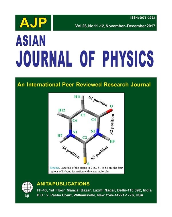
2-Thiouracil: Influence of water in the first hydration shell and Effect of the
Sulfur Atom on the Base Pairs 2-Thiouridine-Adenosine
M Alcolea Palafox1,V K Rastogi2,3, S P Singh4 and S K Rathor3
1Departamento de Química-Fisica1. Facultad de Ciencias Químicas. Universidad Complutense. Madrid- 28040. Spain.
2R D Foundation Group of Institutions, NH-58, Kadrabad, Modinagar (Ghaziabad), India
3Indian Spectroscopy Society, KC 68/1, Old Kavinagar, Ghaziabad-201 002, India
4Department of Physics, Dr B R Ambedkar Govt Degree College, Mainpuri, India
The crystal unit cell of 2-thiouracil (2TU) in the solid state was simulated through a tetramer form using DFT methods. The first and second hydration shells were simulated by explicit number of water molecules surrounding 2TU up to 30. The calculated spectra were compared to the experimental ones. A linear scaling procedure (LSE) was used for this task. The effect of the hydration on different parameters of the molecular structure of 2TU was analyzed. The total atomic charges were discussed. The effect of the sulfur atom on the Watson-Crick (WC) and reverse WC base pair uridine-adenosine was estimated, and the CP corrected interaction energies were calculated. A microhelix RNA:DNA was simulated with two nucleotides base pairs © Anita Publications. All rights reserved.
Keywords: 2-thiouracil, interaction energies, DFT, hydration, scaling, 2-thiouridine, uridine.
Total Refs : 37
| Asian Journal of Physics | Vol. 26 No 11 & 12 (2017) 353-357 |
Electro-optic investigations on ferroelectric and silica nanoparticles doped ferroelectric liquid crystal mixtures
Rajbir Singh
1Department of Physics, Meerut College, Meerut-250 001, India
Dispersed liquid crystal composites have attracted significant interest among scientific community due to their practical and technological applications in various fields including displays. In this work, a ferroelectric liquid crystal and silica nanoparticles doped ferroelectric liquid crystal composites are studied in SmC* phase. The effect of doping and temperature on the spontaneous polarization, switching time and viscosity of FLC are investigated in thin planar sample cell of thickness 9 µm. The doping of silica decreases the polarization and increases the switching time. The viscosity of the sample also changes after dispersion of silica nanoparticles. © Anita Publications. All rights reserved.
Keywords: Ferroelectric liquid crystal, Silica nanoparticle, Polarization, Switching time.
References
- Bellini T, Clark N A, Muzny C D, Phys Rev Lett, 69(1992)788; doi.org/10.1103/PhysRevLett.69.788.
- Bellini T, Clark N A, Schaefer D W, Phys Rev Lett, 74, 2740, (1995)..
- Malik P, Chaudhary A, Raina K K, Asian J Chem, 21(2009)S095–S098.
- Kreuzer M, Tschudi T, Eidenschink R, Mol Cryst Liq Cryst, 223(1992)219–227.
- Kreuzer M, Tschudi T, Jeu W H D, Eidenschink R, Appl Phys Lett, 62(1993)1712; https://doi.org/10.1063/1.109582.
- Rozanski S A, Thoen J, J Non-Cryst Sol, 33-36(2005)2802-2808.
- Kutnjak Z, Kralj S, Zumer S, Phys Rev E, 66, 041702 (2002); doi.org/10.1103/PhysRevE.66.041702.
- Qi H, Hegmann T, J Mater Chem, 18(2008)3288–3294.
- Reznikov Y, Buchnev O, Tereshchenko O, Reshetnyak V, Glushchenko A, Appl Phys Lett, 82(2003)1917; doi.org/10.1063/1.1560871.
- Reshetnyak V Y, Mol Cryst Liq Cryst, 421(2004)219–224,
- Kaur S, Singh S P, Birader A M, Choudhary A, Sreenivas K, Appl Phys Lett, 91, 0231201 (2007); doi.org/10.1063/1.2756136.
- Liang H H, Xiao Y Z, Hsh F J, Wu C C, Lee J Y, Liq Cryst, 37(2010)255–261.
- Jakli A, Almsay L, Borbely S, Rosta L, Eur Phys J, 10(1999)509–513.
- Glushchenko A V, Kresse H, Reznikov Y, Yaroshchuk O, Proc SPIE, 2795, 38 (1996); doi.org/10.1117/12.239228.
- Prasad S K, Sandya K L, Nair G G, Hiremath U S, Yelmaggad C V, Sampth S, Liq Cryst, 33(2006)1121–1125.
- Mertelj A, Jakli A, Copic M, Mol Cryst Liq Cryst, 331(1999)81–87.
- Kingston Chemical, UK.
- Sigma Aldrich, INDIA.
- Neeraj, K. K. Raina, Phase Transitions, 83(2010)615–626.
- Malik P, Raina K K, Bubnov A, Prakash C, Phase Transitions, 79 (2006)889–898
| Asian Journal of Physics | Vol. 26, Nos 11 & 12 (2017) 359-364 |
Effect of the presence of 5- Guanidino-4- nitroimidazole on B- DNA structure
Neena Srivastavaa, A Dwivedib, P K Tripathic and K Singhd
aDepartment of Chemistry, Mahila Vidyalaya Degree College, Aminabad, Lucknow-226 018, India
bChemistry Section, Directorate of Geology and Mining U.P., Lucknow-226 001, India
cFood Safety and Drug Administration, Collectorate Campus, Bahjoi (Sambhal)- 244 410, India
dDepartment of Molecular Microbiology and Immunology, Bond Life Science Centre,
University of Missouri, Columbia, MO 65211, USA
5-Guanidino-4-nitroimidazole (GN), derived from the oxidation of guanine by reactive oxygen and nitrogen species, contains an unusual flexible ring-opened structure. In this molecule, the nitro and guanidino groups possess multiple hydrogen bonding capabilities. In vitro primer extension experiments with bacterial and mammalian polymerases have shown that it is incorporated against C as well as A and G, depending on the polymerase. To elucidate structural and thermodynamic properties of the mutagenic GN lesion, we have investigated the structure of the modified base itself and the GN-containing nucleoside with semi-empirical quantum mechanical calculations at PM3 level and have used molecular modelling techniques (SYBYL) to determine its status in B-DNA duplexes, with four partner bases opposite the GN. Our results show that GN adopts a planar structure at the damaged base level. However, in the nucleoside and in DNA duplexes, steric hinderance between the guanidino group and its linked sugar causes GN to be nonplanar. The GN lesion can adopt both syn and anti conformations on the DNA duplex level, with the guanidino group positioned in the DNA major and minor grooves, respectively. On the basis of hydrogen bonding and stacking interactions, groove dimensions, and bending, we noted that the least distorted GN-modified duplex contains partner C. However, hydrogen bonding interactions between GN and partner G or A are also found, which is similar to the geometry as that observed for mismatches.© Anita Publications. All rights reserved.
Keywords: Ferroelectric liquid crystal, Silica nanoparticle, Polarization, Switching time.
Peer Review Information
Method: Single- anonymous; Screened for Plagiarism? Yes
Buy this Article in Print © Anita Publications. All rights reserved
References
- Inoue M, Sato E F, Nishikawa M, Park A M, Kira Y, Imada I, Utsumi K, Mitochondrial generation of reactive oxygen species and its role in aerobic life, Curr Med Chem,10(2003)2495-2505.
- Le Bras M, Clement M V, Pervaiz S, Brenner C, Reactive oxygen species and the mitochondrial signaling pathway of cell death, Histol Histopathol, 20(2005)205-219.
- Cadet J, Berger M, Douki T, Ravanat J L, Oxidative damage to DNA: formation, measurement, and biological significance, Rev Physiol Biochem Pharmacol, 131(1997)1-87.
- Dizdaroglu M, Chemical determination of free radical-induced damage to DNA, Free Radical Biol Med, 10(1991) 225-242.
- Epe B, DNA Damage Profiles Induced by Oxidizing Agents, Rev Physiol Biochem Pharmacol, 127(1995)223-249.
- Lindahl T, Instability and decay of the primary structure of DNA, Nature, 362(1993)709-715.
- Greenberg M M, In vitro and in vivo effects of oxidative damage to deoxyguanosine, Biochem Soc Trans, 32(2004) 46-50.
- Hussain S P, Hofseth L J, Harris C C, Radical causes of cancer, Nat Rev Cancer, 3(2003)276-285.
- Klaunig J E, Kamendulis L M, The role of oxidative stress in carcinogenesis, Annu Rev Pharmacol Toxicol, 44(2004)239-267.
- Olinski R, Gackowski D, Foksinski M, Rozalski R, Roszkowski K, Jaruga P, Oxidative DNA damage: assessment of the role in carcinogenesis, atherosclerosis, and acquired immunodeficiency syndrome, Free Radical Biol Med, 33(2002)192-200.
- Weinberg R A, How cancer arises, Sci Am, 275(1996)62-70.
- Finkel T, Holbrook N J, Oxidants, oxidative stress and the biology of ageing, Nature, 408(2000)239-247.
- Mandavilli B S, Santos J H, Van Houten B, Mitochondrial DNA repair and aging, Mutat Res, 509(2002)127-151.
- Sastre J, Pallardo F V, Vina J, The role of mitochondrial oxidative stress in aging, Free Radical Biol Med, 35(2003)1-8.
- Hamilton M L, Van Remmen H, Drake J A, Yang H, Guo Z M, Kewitt K, Walter C A, Richardson A, Does oxidative damage to DNA increase with age?, Proc Natl Acad Sci (USA), 98(2001)10469-10474.
- Osterod M, Hollenbach S, Hengstler J G, Barnes D E, Lindahl T, Epe B, Age-related and tissue-specific accumulation of oxidative DNA base damage in 7,8-dihydro-8-oxoguanine-DNA glycosylase (Ogg1) deficient mice, Carcinogenesis, 22(2001)1459-1463.
- Cooke M S, Evans M D, Dizdaroglu M, Lunec J, Oxidative DNA damage: mechanisms, mutation, and disease, FASEB J, 17(2003)1195-1214.
- Neeley W L, Delaney J C, Henderson P T, Essigmann J M, In vivo bypass efficiencies and mutational signatures of the guanine oxidation products 2-aminoimidazolone and 5-guanidino-4-nitroimidazole, J Biol Chem, 279(2004)43568-43573.
- Gu F, Stillwell W G, Wishnok J S, Shallop A J, Jones R A, Tannenbaum S R, Peroxynitrite-induced reactions of synthetic oligo 2′-deoxynucleotides and DNA containing guanine: formation and stability of a 5-guanidino-4-nitroimidazole lesion, Biochemistry, 41(2002)7508-7518.
- Stewart J J P, Optimization of parameters for semiempirical methods I. Method, J Comput Chem, 1989, 10(1989)209-220.
[Received: 29.01.2017; revised recd: 01.06.2017; accepted: 01.08.2017]
| Asian Journal of Physics | Vol. 26, Nos 11 & 12 (2017) 365-373 |
Molecular structure and vibrational spectra of 2-thiouracil: A comparison with uracil
Kaushal Rani1,2, Jay Prakash3, S P Singh4, J K Vats5, M A Palafox6 and V K Rastogi1
1Indian Spectroscopy Society, KC-68/1, Old Kavinagar, Ghaziabad-201 002, India
2Department of Physics, Meerut College, Meerut-250 003, India
3Department of Physics, Kamla Nehru P.G. College, Tej Gaon, Raebareli-229 215, India
4Department of Physics, Dr B R Ambedkar Govt Degree College, Mainpuri-205 263, India
5P G Department of Physics, Jai Prakash University, Chapra-841 301, India
6Departamento de Química-Física, Facultad de C Químicas, Universidad Complutense, Madrid-28040, Spain.
Spectroscopic and structural studies of uracil and its derivatives have been reported both theoretically and as well experimentally by many authors. Uracil and its thio analogue 2-thiouracil (2-TU) affect the growth of plants. To understand biochemically the mode of inhibitory or malforming actions of uracil and 2-TU on the growth of rice and wheat plants, the knowledge of their structures is essential. Hence, in the present work an attempt has been made to study their spectra and structures using vibrational spectroscopy as technique. The replacement of an oxygen atom by sulphur leads to a shift of the experimental ν(N-H) bands to lower wavenumbers, 27 cm–1 for the N1-H mode and 20 cm–1 for N3-H. The effect of sulphur substitution on ν(N3-H) wavenumber and intensity reflects changes in proton abilities of this group as well as the hydrogen bonding in which they participate. Compared to uracil, the sulfur atom in 2-TU results mainly in a significant change of the bond-length at the substitution site: S=C ~1.66 Å, as compared to C=O ~1.22 Å. This fact leads to a slightly reduction in the neighboring bond lengths N1-C2 and N3-C2, but the N-H and C-H bonds are little affected. © Anita Publications. All rights reserved.
Keywords: Uracil, 2-Thiouracil, FT-IR, FT-Raman, Density Functional Theory (DFT)
References
- Palafox M Alcolea, Iza N, Gil M, The hydration effect on the uracil frequencies: an experimental and quantum chemical study, J Molec Struct (Theochem), 585(2002)69–92.
- Palafox M A, Rastogi V K, Quantum chemical predictions of the vibrational spectra of polyatomic molecules.The uracil molecule and two derivatives, Spectrochim Acta, A58(2002)411–440.
- Palafox M A, Rastogi V K, Tanwar R P, Mittal L,Vibrational frequencies and structure of 2-thiouracil by Hartree-Fock, post Hartree-Fock and density functional methods, Spectrochim Acta, A59(2003)2473–2486.
- Kattan D, Palafox M A, Rathor S K, Rastogi V K, A DFT analysis of the molecular structure, vibrational spectra and other molecular properties of 5-nitrouracil and comparison with uracil, J Mole Struct, 1106(2016)300–315.
- Mathur S N, Sharma R A, Effect of Uracil and 5-Nitrouracil on Growth and Flowering of Tomato, Physiology Plantarum, 21(1968)911; doi.org/10.1111/j.1399-3054.1968.tb07317.x..
- Turan Y, Konuk M, The Effect of Uracil on the Germination and Growth of some leguminous Plants, Tr J Botany, 23(1999)241–244.
- Inouye J, Jun-Ichiro M J, Effect of 2-thiouracil on flower initiation in rice and wheat plants grown under aseptic conditions, J Faculty Agriculture, Kyushu University, 14(1966)33–41.
- Da Silva M A R, Amaral L M, Szterner P, Experimental study on the thermochemistry of 2-thiouracil, 5-methyl-2-thiouracil and 6-methyl-2-thiouracil, J Chem Thermodyn, 57(2013)380–386.
- Harsányi L, Császár P, Császár A, Boggs J E, Interpretation of the vibrational spectra of matrix-isolated uracil from scaled ab initio quantum mechanical force fields, Int J Quantum Chem, 29(1986)799–815.
- Barnes A J, Stuckey M A, Gall L Le, Nucleic acid bases studied by matrix isolation vibrational spectroscopy: Uracil and deuterated uracils, Spectrochim Acta, A40(1984)419–431.
- Wojcik M J, Medium-frequency Raman spectra of crystalline uracil, thymine and their 1-methyl derivatives, J Mole Struct, 219(1990)305–310.
- Singh R, Jaiswal S, Kumar M, Singh P, Srivastav G, Yadav R A, DFT study of molecular geometries and vibrational characteristics of uracil and its thio-derivatives and their radical cations, Spectrochim Acta, A75(2010)267–276.
- Grosmaire L, Delarbre J-L, Vibrational spectra of 6-methyluracil, 6-methyl-2-thiouracil and their deuterated analogues, J Mole Struct, 1011(2012)42–49.
- Gnanasambandan T, Gunasekaran S, Seshadri S, Vibrational spectroscopic investigation on propylthiouraci, Int J Recent Scientific Res, 3(2012)590–597.
- Rostkowska H, Barski A, Szczepaniak K, Szczesniak M, Person W B, The tautomeric equilibria of thioanalogues of nucleic acids: spectroscopic studies of 2-thiouracils in the vapour phase and in low temperature matrices, J Mole Struct, 176(1988)137–147.
- Rostkowska H, Szczepaniak K, Nowak M J, Leszczynski J, KuBulat K, Person W B, Thiouracils. 2. Tautomerism and infrared-spectra of thiouracils. Matrix-isolation and ab initio studies, J Am Chem Soc, 112(1990)2147–2160.
- Graindourze M, Grootaers T, Smets J, Zeegers-Huyskens Th, Maes G, FT-IR spectroscopic study of uracil derivatives and their hydrogen bonded complexes with proton donors: II. Monomer IR absorptions of thiouracils and 5-halogeno- uracils in argon matrices, J Mole Struct, 237(1990)389–410.
- Lapinski L, Rostkowska H, Nowak M J, Kwiatkowski J S, Leszczynski J, Infrared spectra of thiouracils: Experimental matrix isolation and ab initio Hartree-Fock, post-Hartree-Fock and density functional theory studies Vib Spectrosc, 13(1996)23–40.
- Yadav R A, Yadav P N S, Yadav J S, Vibrational studies of biomolecules. 1. 2-Thiouracil, Proc Indian Acad Sci, (Chem Sci), 100(1988)69–78.
- Seminario J M, Politzer P (eds).Modern Density Modern Density Function Theory, A Tool for Chemistry, (Elsevier), 1995.
- Gaussian 09, Revision D.01, Frisch M J, Trucks G W, Schlegel H B, Scuseria G E, Robb M A, Cheeseman J R, Scalmani G, Barone V, Mennucci B, Petersson G A, Nakatsuji H, Caricato M, Li X, Hratchian H P, Izmaylov A F, Bloino J, Zheng G, Sonnenberg J L, Hada M, Ehara M, Toyota K, Fukuda R, Hasegawa J, Ishida M, Nakajima T, Honda Y, Kitao O, Nakai H, Vreven T, Montgomery J A, (Jr), Peralta J E, Ogliaro F, Bearpark M, Heyd J J, Brothers E, Kudin K N, Staroverov V N, Kobayashi R, Normand J, Raghavachari K, Rendell A, Burant J C, Iyengar S S, Tomasi J, Cossi M, Rega N, Millam J M, Klene M, Knox J E, Cross J B, Bakken V, Adamo C, Jaramillo J, Gomperts R, Stratmann R E, Yazyev O, Austin A J, Cammi R, Pomelli C, Ochterski J W, Martin R L, Morokuma K, Zakrzewski V G, Voth G A, Salvador P, Dannenberg J J, Dapprich S, Daniels A D, Ö. Farkas, Foresman J B, Ortiz J V, Cioslowski J, Fox D J, Gaussian, Inc., Wallingford CT, 2009.
- Palafox M Alcolea, Talaya J, Guerrero-Martínez A, Tardajos G, Kumar H, Vats J K, Rastogi V K, Quantum chemical scaling and its importance: The infrared and Raman spectra of 5-bromouracil, Spectrosc Lett, 43(2010)51–59.
- Bourova T, Ten G, Andreeva S, Berezin V, Theoretical analysis of Raman and resonance Raman spectra of simple bases of nucleic acids, J Raman Spectrosc, 31(2000)827–836.
- Palafox M Alcolea, Rastogi V K, Guerrero-Martínez A, Tardajos G, Joe H, Vats J K, Simulation of a tetramer form of 5-iodouracil: The vibrational spectra and molecular structure in the isolated and in the solid state by using DFT calculations, Vib Spectrosc, 52(2010)108–121.
- Palafox M Alcolea, Tardajos G, Guerrero-Martínez A, Rastogi V K, Mishra D, Ojha S P, Kiefer W, FT-IR, FT-Raman spectra, density functional computations of the vibrational spectra and molecular geometry of biomolecule 5-aminouracil, Chem Phys, 340(2007)17–31.
- Rastogi V K, Jain V, Yadav R A, Singh C, Palafox M Alcolea, Fourier transform Raman spectrum and ab initio and density functional computations of the vibrational spectrum, molecular geometry, atomic charges and some molecular properties of the anticarcinogenic drug 5-fluorouracil,J Raman Spectrosc, 31(2000)595–603.
- Rastogi V K, Palafox M Alcolea, Mittal L, Peica N, Kiefer W, Lang K, Ojha S P, FTIR and FT-Raman spectra and density functional computations of the vibrational spectra, molecular geometry and atomic charges of the biomolecule: 5-bromouracil, J Raman Spectrosc. 38(2007)1227–1241.
- Palafox M Alcolea, Tardajos G, Guerrero-Martínez A, Vats J K, Joe H, Rastogi V K, Relationships observed in the structure and spectra of uracil and its 5-substituted derivatives, Spectrochim Acta, A75(2010)1261–1269.
- Palafox M Alcolea, Nielsen O F, Lang K, Garg P, Rastogi V K, Geometry and vibrational spectra of 5-substituted uracils, Asian Chem Lett, 8(2004)81–93.
- Rastogi V K, Singh C, Jain V, Palafox M Alcolea, FTIR and FT-Raman spectra of 5-methyluracil (thymine), J Raman Spectrosc, 31(2000)1005–1012.
- Palafox M Alcolea, Rastogi V K, Density functional theory computations on vibrational spectra: Scaling procedures to improve the results, Asian J Phys, 20(2011)103–121.
- Palafox M Alcolea, Rastogi V K, Singh S P, Rathor S, 2-Thiouracil: Influence of water in the first hydration shell and Effect of the Sulfur Atom on the Base Pairs 2-Thiouridine-Adenosine, Asian J Phys, 26(2017)337–351.
- Palafox M A, Rastogi V K, 6-Aminouracil: Geometries and spectra in the isolated state and in the solid state simulation. A comparison with 5-aminouracil, J Mole Struct, 1108(2016)482–495.
- Palafox M A, Kumar H, Sharma M, Joe H, Rastogi V K, Spectra and Structure of Uracil and its 5-Haloderivatives: A Review, Advancements and Futuristic Trends in Material Science, Eds. Khan M S, Gupta A,(Allied Publishers, New Delhi), 2011, pp 70-87.