Editor-in-Chief : V.K. Rastogi

ASIAN JOURNAL OF PHYSICS
An International Peer Reviewed Research Journal
Frequency : Monthly,
ISSN : 0971 – 3093
Editor-In-Chief (Hon.) :
Dr. V.K. Rastogi
e-mail:[email protected]
[email protected]
| AJP | ISSN : 0971 – 3093 Vol 28, No 2, February, 2019 |
Asian
Journal of Physics
|
Asian Journal of Physics |
Vol. 28 No 2 (2019) 71-93 |
Vibrational spectroscopic, molecular docking and in-vitro cytotoxicity studies against lung cancer cell line on 5-cyano-indole-3-carboxaldehyde
S Christopher Jeyaseelan1, R Premkumar1, K Kaviyarasu2, 3, and A Milton Franklin Benial1
1PG and Research Department of Physics, N M S S VN College, Madurai-625 019, Tamil Nadu, India.
2UNESCO-UNISA Africa Chair in Nanosciences/Nanotechnology Laboratories,
College of Graduate Studies, University of South Africa (UNISA), South Africa
3Nanosciences African network (NANOAFNET), Materials Research Department (MRD), iThemba LABS-National Research Foundation (NRF),
1 Old Faure Road, 7129, P O Box 722, Somerset West, Western Cape Province, South Africa.
The most stable and optimized molecular structure of 5-cyano-indole-3-carboxaldehyde (CICA) molecule was predicted by density functional theory using B3LYP method with cc-pVTZ basis set. The calculated and experimentally observed FT-IR and FT-Raman vibrational frequencies were assigned and compared. Ultraviolet-visible spectrum of the molecule was simulated and validated experimentally. Molecular docking analysis of CICA molecule forms a stable complex with mGluRs targeted protein which is evident from the predicted binding energy values and hence it can act as an effective anti-Parkinson agent. The molecular reactivity, ionization energy, electron affinity and other relative molecular properties of the molecule were studied using frontier molecular orbitals analysis. Mulliken atomic charge distribution and Fukui functions analyses were performed to predict the reactive sites within the molecule. The molecular electrostatic potential surface was simulated to predict the possible electrophilic and nucleophilic reactive sites of the molecule. Natural bond orbital (NBO) analysis gives the information about intra- and intermolecular interactions of the molecule. NBO analysis indicates the higher stabilization interaction between LP orbital and π*/σ* orbital, which is responsible for the bioactivity of the molecule. The in-vitro cytotoxicity study of title molecule was performed using MTT assay technique which reveals that CICA molecule can act as a potential inhibitor against human pulmonary epithelial lung cancer cell line (A549).
Keywords: FT- IR, FT-Raman, 5-cyano-indole-3-carboxaldehyde, Molecular docking, Anti-Parkinson agent, MTT assay.
Total Refs : 59
|
Asian Journal of Physics |
Vol. 28 No 2 (2019) 95-104 |
Infrared, Raman and surface-enhanced Raman scattering spectra of sarcosine
Ying-Sing Lia*, JingciaChengb and Yu Wangb
aDepartment of Chemistry, University of Memphis, Memphis, TN 38138 USA
bWuxi JC Pharmaceutical Technology, Wuxi, Jiangsu 214036 China
Mid-infrared and Raman spectra of sarcosine in solid state and in solution have been recorded. Results agree with the expectation that sarcosine exists as zwitterionic form in solid and neutral aqueous solution. Vibrational assignments have been suggested based on the relative spectral intensities alongwith the group frequencies and related references. Surface-Enhanced Raman Scattering (SERS) spectrum of sarcosine adsorbed on colloidal nano particles has been recorded. Silver coated iron oxide magnetic nano-particles (MNPs) have been prepared and used as substrate for recording SERS spectra of sarcosine. The interpretation of the SERS spectral bands was given in reference to the results of vibrational assignments. Results obtained from the present study should be helpful in initiating a pathway for the trace analysis of sarcosine by using SERS spectroscopy. © Anita Publications. All rights reserved..
Keywords: Sarcosine, Infrared spectra, Raman spectra, Surface-Enhanced Raman Scattering, Magnetic nano-particles.
References
1. Mostad A, Natarajan S, Acta Chem Scand, 43(1989)1004-1006.
2. Trzebiatowska-Gusowska M, Gagor A, Acta Cryst Sec E, 63(2007)4694-4694.
3. Saha N N, Mazumdar S K, Bhattacharyya S C, Sci Cult, 34(1968)47-49.
4. Bhattacharyya S C, Saha N N, J Cryst Mol Struct, 8(1978)105-113.
5. Sannie C, Poremski V, Bull Soc Chem France, 8(1941)702-713.
6. Edsall J T, J Am Chem Soc, 65(1943)1767-1770.
7. Novak A, Cotrait M, Ann Chim, 1(1966)263-270.
8. Stahlberg U, Steger E, Spectrochim Acta, A 23(1967)475-490.
9. Cocinero E J, Villanueva P, Lesarri A, Sanz M E, Blanco S, Mata S, Lopez J C, Alonso J L, Chem Phys Lett, 435(2007)336-341.
10. Gomez-Zavaglia A, Fausto R, Vib Spectrosc, 33(2003)105-126.
11. Sreekumar A, Poisson L M, Rajendiran T M, Khan A P, Cao Q, Yu J D, Laxman B, Mehra R, Lonigro R J, Li Y, Nyati M K, Ahsan A, Kalyana-Sundaram S, Han B, Cao X H, Byun J, Omenn G S, Ghosh D, Pennathur S, Alexander D C, Berger A, Shuster J R, Wei J T, Varambally S, Beecher C, Chinnaiyan A M, Nature, 457(2009)910-914.
12. Cernei N, Heger Z, Gumulec J, Zitka O, Masarik M, Babula P, Eckschlager T, Stiborova M, Kizek R, Adam V, Int J Mol Sci, 14(2013)13893-13908.
13. Jentzmik F, Stephan C, Miller K, Schrader M, Erbersdobler A, Kristiansen G, Lein M, Jung K, Eur Urol, 58(2010)12-18.
14. Struys E A, Heijboer A C, Moorselaar J V, Jakobs C, Blankenstein M A, Ann Clin Biochem, 47(2010)282-282.
15. Pavlou M, Diamandis E P, Clin Chem, 55(2009)1277-1279.
16. Li Y S, Cheng J C, Coons L B, Spectrochim Acta, 55(1999)1197-1207.
17. Li Y S, Church J S, Woodhead A L, J Magn Magn Mat, 324(2012)1543-1550.
18. Li Y S, Wang Y, Appl Spectrosc, 46(1992)142-146.
19. Kim K, Jang H J, Shin K S, Analyst, 134(2009)308-313.
20. Parameswari A, Asath R M, Premkumar R, Benial A M F, J Mol Struct, 1138(2017)102-109.
21. Stepanian S G, Reva I D, Radchenko E D, Rosado M T S, Duarte M, Fausto R, Adamowicz L, J Phys Chem A, 1102(998)1041-1054.
22. Gomez-Zavaglia A, Fausto R, Phys Chem Chem Phys, 5(2003)3154-3161.
23. Lin-Vien D, Colthup N B, Gateley W G, Grasselli J G, The Handbook of Infrared and Raman Characteristic Frequencies of Organic Molecules, (Academic Press: New York, USA), 1991.
24. Kirschenbaum D M, Appl Spectrosc, 17(1963)149-153.
25. Inomata Y, Shibata A, Yukawa Y, Takeuchi T, Moriwaki T, Spectrochim Acta, 44(1988)97-107.
26. Ghazaaryan V V, Fleck M, Petrosyan A M, J Mole Struct, 121(2012)130-137.
27. Socrates G, Infrared and Raman Characteristic Group Frequencies, 3rd edn, (John Wiley & Sons: New York, USA), 2001.
28. Bellamy LJ, The Infra-red Spectra of Complex Molecules, 3rd. edn, (Chapman and Hall: London, GB), 1975.
29. Colthup N B, Daly L H, Wiberley S E, Introduction to Infrared and Raman Spectroscopy, 3rd edn, (Academic Press: Boston, USA), 1990.
30. Ding Y B, Kroghjespersen K, Chem Phys Lett, 199(1992)261-266.
31. Suh J S, Moskovits M, J Am Chem Soc, 108(1986)4711-4718.
32. Fang JH, Yuan L, Jin YY, Li HB, Yang HJ, Proc. Soc. Photo-Optic. Instr. Eng. , Beijing, China, Oct. 8, 2004; Zhu X, Chou S Y, Arakawa Y. Eds. SPIE (2005)468-472.
33. Stewart S, Fredericks P M, Spectrochim Acta, 55 A(1999)1641-1660.
34. Acharya P K, Narayanan P S, J Raman Spectrosc,1(1973)499-505.
35. Suh J S, Dillela D P, Moskovits M J, J Phys Chem, 87(1983)1540-1544.
36. Sharma P, Singh D K, Gupta V, Asthana B P, Mishra P C, Singh R K, Spectrochim Acta,116 A(2013)74-83.
37. Li Y S, Church J S, Woodhead A L, Moussa F, Spectrochim Acta, 76A(2010)484-489.
38. Pang Y F, Wang C W, Wang J, Sun Z W, Xiao R, Wang S Q, Biosen Bioelec, 79(2016)574-580.
39. Toma S H, Santos J J, Araki K, Toma H E, Anal Chim Acta, 855(2015)70-75.
40. Bhana S, Wang Y M, Huang X H, Nanomed, 10(2015)1973-1990.
|
Asian Journal of Physics |
Vol. 28 No 2 (2019) 143-148 |
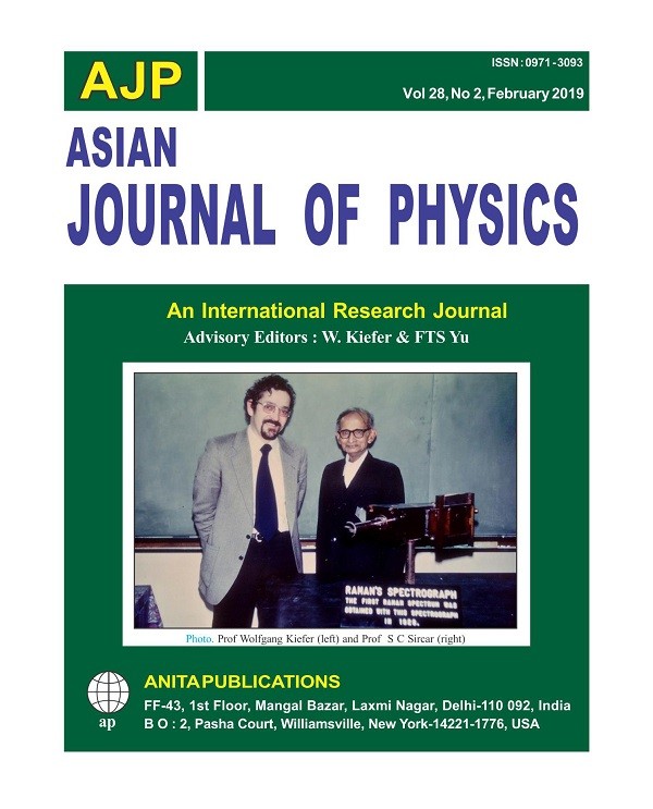
S C Sirkar – The witness of the discovery of the Raman effect
Rajinder Singh
Research Group – Physics Didactic and History of Science
Physics Institute, University of Oldenburg, Germany
On Feb 28, 1928, the Raman effect was discovered by C V Raman and his students. Raman received the Nobel Prize in 1930 for his work on light scattering and the effect named after him. Russian physicists, who discovered the same effect nearly at the same time, were ignored. These details are explored in the previous two articles published in “Asian J Physics” [1,2].
According to the record of the Nobel Foundation, Raman is the only one Indian Nobel Laureate in the field of Physics. Not surprisingly there are a number of biographies, which deal with various aspects of his life [3-14]. However, to the best of my knowledge, none of the authors has discussed Sukumar Chandra Sirkar’s view about the discovery of Raman Effect. In those days S C Sirkar (Photo 1) was Raman’s research scholar at the University of Calcutta. The present article is intended to fulfill the gap.
S C Sirkar – The witness of the discovery of the Raman effect.pdf
Rajinder Singh