Editor-in-Chief : V.K. Rastogi


ASIAN JOURNAL OF PHYSICS
An International Peer Reviewed Research Journal
Frequency : Monthly,
ISSN : 0971 – 3093
Editor-In-Chief (Hon.) :
Dr. V.K. Rastogi
e-mail:[email protected]
[email protected]
| AJP | ISSN : 0971 – 3093 Vol 31, No 2, Feb 2022 |
Asian
Journal of Physics
Vol 31 No 2 February 2022
A Special Issue
on
Women in Science: Raman Spectroscopy
 Guest Editors : Giulietta Smulevich & Wolfgang Kiefer
![]()
ANITA PUBLICATIONS
FF-43, 1st Floor, Mangal Bazar, Laxmi Nagar, Delhi-110 092, India
Asian Journal of Physics
(A Publication Not for Profit)
Volume 31, No 2 (2022)
CONTENTS |
Guest Editorial
About the Guest Editors
Message
My career path in the field of Raman spectroscopy and microscopy
Fran Adar177
Biopolymers in the eggshell of the Argentine Black and White Tegu (Salvator merianae): a Raman microscopy study
Rosa María Susana Álvarez, Alfredo Nicolás Dominguez, Francisco Alejandro Cortez, Oscar Augusto Carlino-Aráoz, and Fernando Horacio Campos-Casal195
Infrared active vibrations in doped π-conjugated materials: the mechanism of activation of Raman modes
C Castiglioni and M Tommasini211
Ultrafast vibrational and electronic relaxation of carotenoids investigated with Femtosecond Stimulated Raman Spectroscopy
E Ragnoni, T M Kardaś, A Lapini, P Foggi, R Righini and M Di Donato227
The role of Raman spectroscopy in food and beverage analysis
Karen Esmonde-White and Mary Lewis 241
Raman and infrared microscopic study on the lipid redistribution in Alzheimer diseased murine tissue
Alicia Schirer, Youssef El Khoury, Christine Patte-Mensah, Christian Klein, Laurence Meyer, Monika Rataj-Baniowska, David Moss, Ayikoe-Guy Mensah-Nyagan Sophie Lecomte and Petra Hellwig 259
The use of thin layer chromatography combined with surface-enhanced Raman spectroscopy for the identification of controlled substances
Kasey R Cargill, Marisia A Fikiet, and Brooke W Kammrath 265
A new generation of Raman spectroscopists: Thinking diversity in the analytical sciences
Janina Kneipp, Ulrich Panne, Dorota Bartkowiak, Dimitra Gkogkou, Christian Heck, Wan-Ing Lin, Tilmann Neubert, Elena Pavlenko, Christine Joy Uy Querebillo, Bita Rezania, Maximilian Ries, Victor M Rodriguez Zancajo, Radwan Mohammed Sarhan, Gergo Péter Szekeres, Yanlong Xin, Anur Yadav, Zhiyang Zhang and Vesna Zivanovic 283
Detection of immune reaction by surface-enhanced Raman Spectroscopy: A mini-review
Kamilla Malek and Ewelina Wiercigroch303
Raman spectroscopy in multidisciplinary approaches applied to drug design
Maria Paula M Marques315
Establishing the SERS-based sensing capabilities of silver nanorod thin films fabricated through oblique angle deposition at different temperatures
Adam C Stahler, Piyush J Shah, Andrew M Sarangan, and Ioana E Pavel 341
Drug-drug interaction: the case of flubendazole and doxycycline hyclate investigated by Raman spectroscopy
Ilirjana Bajama, Luisa Andronie, Simona Cinta Pinzaru355
Bromide hydration as a function of concentration and temperature
Letizia Scarabattoli, Lucia Comez and Paola Sassi365
Raman capability to study heme proteins
Giulietta Smulevich375
The characteristics of the carbon nanotubes layer deposited by the electrophoretic method on a titanium substrate studied by Raman micro-spectroscopy and nanoindentation
Maria Pajda, Aleksandra Wesełucha-Birczyńska, Sylvia Turrell, Aleksandra Benko and Marta Blażewicz391
Guest Editorial
Professor Vinod K Rastogi, Chief-Editor of the Asian Journal of Physics, has kindly invited us to be Guest-Editors for a Special Issue entitled “Women in Science: Raman Spectroscopy”, which we immediately accepted with great pleasure. Before we summarize in chapter 3) the accepted contributions, we like to briefly describe 1) C V Raman’s attitude towards women scientists and 2) the early (1969 – 1978) recognition of women scientists in laser Raman spectroscopy and hereby demonstrate how this has been considerably changed in the last half century.
1) C V Raman’s attitude towards women scientists
When one of us (WK) had correspondence with Fran Adar, author of the first paper in this issue, she pointed to the name of Kamala Sohonie, who, according to Wikipedia [1] became the first woman who had been accepted into and worked at the Indian Institute of Science (IISc), Bangalore, India. However, when she first applied to IISC for a research fellowship “her application was turned down by the then-Director Prof C V Raman on the grounds that women were not considered competent enough to pursue research” [2]. According to Ref [1], she was granted permission only under very strict conditions: (i) being on probation for the first year, (ii) working only at late night, (iii) not spoiling the environment of the lab (not being a ‘distraction’ to the male researchers). Her dedication and research mettle influenced Prof Raman’s decision to let women into the IISc a year after she completed her M Sc degree with distinction in 1936. Many women got their admission to the institution [2].
We are sure that Raman would have been most happy knowing about the tremendous scientific work women have contributed meanwhile to the research area for which he had been the pioneer.
2) Early (1969-1978) recognition of women scientists in laser Raman spectroscopy
As well known, the practice of Raman spectroscopy was completely revolutionized in 1962 when Porto and Wood [3] reported on the first laser excited Raman spectrum using a ruby laser as excitation source. In 1969, the very first International Conference on Raman Spectroscopy (ICORS I) took place in Ottawa, Canada. About 150 scientists attended this meeting, but all of the Invited Lectures had been presented by men only, and there was one woman only who chaired a lecture: Madame Marie-Louise Josien from France, a well-known vibrational spectroscopist at that time (see Fig 1).
Surprisingly, also during the second ICORS meeting in Oxford, England, in 1970, again no woman had been invited for a lecture, despite the fact that about 300 Raman spectroscopists attended this meeting!
The unbalanced recognition of women in Raman spectroscopy slightly improved at the third ICORS, which took place in Reims, France, in 1972. For the first time in the ICORS series one woman (Marie-Louise Josien) out of 15 people had been a member of the Organizing Committee. In addition, the Proceedings [5] of this conference revealed six female authors.
In the Fourth ICORS (Brunswick, Maine, USA) in 1974, out of the 22 Invited Lectures, two were given by female scientists (Monique Constant and Marie-Louise Josien). Also a few contributed lectures had been presented by women, among them one by Fran Adar. Another female speaker, who later on played an important role in industrial applications of Raman spectroscopy, had been Jenny Grasselli from USA.
The Fifth ICORS in 1976 took place in Freiburg, Germany, with a total of 26 Invited Lectures, all given by men. Noteworthy was a contributed paper by Lydia Colombo from former Yugoslavia, who talked on phase transitions in crystals (see Fig 2).
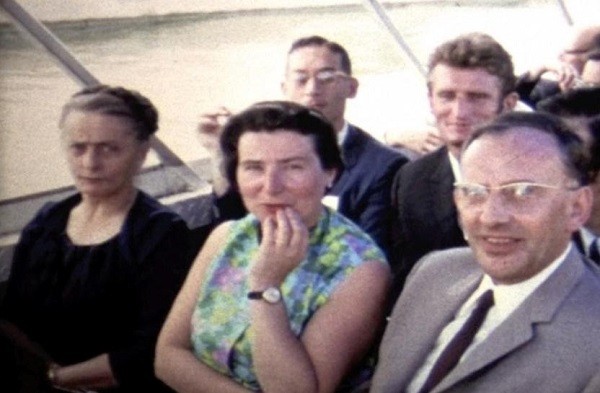
Fig 1. The French Raman Group attending the excursion trip during ICORS I, Ottawa, 1969. From left, front: Madame M L Josien, Mrs M Delhaye, R Dupeyrat, back: J Corset, W Holzer (Germany). Photo extracted from a 2×8 mm movie film taken by W Kiefer. For the movie film itself, see Ref [4].

Fig 2. Lydia Colombo, Zagreb, former Yugoslavia, giving a talk during ICORS V in Freiburg, Germany, 1976. Photo taken by R Claus, Munich.
In 1978, the Raman community celebrated the 50th anniversary of the discovery by C V Raman of the effect which bears his name during the Sixth ICORS in Bangalore, India, the place where Raman spent the last part of his life. Volume 1 of the Proceedings of this meeting [6] reveals that only one women (L Colombo) had been among the Invited 42 Lecturers. During the same ICORS meeting, an International Steering Committee had been founded due to the initiative of Derek A Long, again a group of men only. It took 20 years until a female member (Giulietta Smulevich) was included (in 1996).
Another source for getting information on early recognition of women in Raman Spectroscopy is the Journal of Raman Spectroscopy (JRS) which had been founded in 1975 by Derek A Long and Harold J Bernstein. Among the 50 Associate Editors and Members of the Editorial Board at the beginning, there had been only one women, i.e. Madame Josien. Fortunately, the situation has quite changed since then: instead of 2% female members there are now 20%. We are also happy that the attendance of women in the ICORS meetings has increased considerably since the early days of laser Raman spectroscopy, as example, in the XXV ICORS in Fortaleza, Brazil, 2016, about 10% women (total ca. 400) participated, as derived from the group photo.
3) The contributions from women Raman Spectroscopists in this special issue in alphabetic order
Fran Adar: “My career path in the field of Raman spectroscopy and microscopy”; Horiba Scientific in Edison, New Jersey, USA.
Dr Adar, has spent her scientific life in the field of Raman spectroscopy since the 1960’s. In this review, with a synergy between spectroscopy and materials science, she guides us into an interesting trip in the Raman world from the development of instrumentations to the applications of Raman spectroscopy into materials science. She has collaborated with many scientists around the world, exploring applications in fields that included polymer physics, ceramics, corrosion mechanisms, and pharmaceuticals, spanning her interest from Physics to Biophysics.
Rosa María Susana Álvarez et al: “Biopolymers in the eggshell of the Argentine Black and White Tegu (Salvator merianae): a Raman microscopy study”; Universidad Nacional de Tucumán, Tucumán, Argentina.
The paper by Dr Álvarez et al is focused on the structural analysis of the organic matrix of the Salvator merianae eggshell, carried out by Raman microscopy combined with different microscopy techniques. They report an interesting organization of fibrillar polymers, consisting mainly of different types of collagen and keratins. Raman microscopy evidenced the presence of a gradual change in the relative content between these components at different depth levels of the eggshell cross-section. The spatial distribution of collagen and keratins correlates with the morphology of this complex system.
Chiara Castiglioni and Matteo Tommasini: “Infrared active vibrations in doped π-conjugated materials: the mechanism of activation of Raman modes”; Politecnico di Milano, Milano, Italy.
Drs Castiglioni and Tommasini study the effect of doping polyacetylene with electron acceptor/donor species using Density Functional Theory calculations. The analysis of the predicted spectroscopic observables gives insight into the origin of the strong doping-induced IR vibrational transitions, closely related to the major Raman bands of the undoped species. The computational data indicate that the intramolecular charge hopping is promoted by collective nuclear displacements along the Effective Conjugation Coordinate.
Mariangela Di Donato et al: “Ultrafast vibrational and electronic relaxation of carotenoids investigated with Femtosecond Stimulated Raman Spectroscopy”; LENS (European Laboratory for Non-Linear Spectroscopy and ICCOM-CNR, Sesto Fiorentino (FI), Italy.
In the review by Dr Di Donato et al the photophysics of carotenoids using Femtosecond Stimulated Raman Spectroscopy is presented. After a brief introduction of the technique, the review highlights the potentialities of the technique in disentangling the very fast relaxation processes occurring upon light absorption in carotenoids. The technique allows one to clarify the timescale of vibrational energy relaxation in the electronic excited states and to identify the nature of the excited states which are involved in the photophysics of these pigments, either in solution and when embedded in photosynthetic proteins.
Karen Esmonde-White and Mary Lewis: “The Role of Raman Spectroscopy in Food and Beverage Analysis”; Kaiser Optical Systems, Inc Ann Arbor, MI USA.
Drs Esmonde-White and Lewis present a review on the application of Raman spectroscopy for the analysis of food and beverages. They provide a solid perspective for future application in this field, showing that the vibrational techniques is an established Process Analytical Technology (PAT) for laboratory and within the chemical and life sciences industries. Raman spectroscopy, in fact, can be adopted by the food industry for quality control and storage in the industrial food processing.
Petra Hellwig et al: “Raman and infrared microscopic study on the lipid redistribution in Alzheimer diseased murine tissue”; Université de Strasbourg-CNRS, Strasbourg, France.
Accurate identification of the major structural and molecular changes which spread throughout the cerebral cortex during the neurodegenerative process occurring in the Alzheimer disease (AD) is still of major clinical importance. The review by Dr Hellwig et al presents Raman and infrared microscopic evidence for the reorganization of phospholipids in brain tissue from diseased tissues of mice with severe AD. On the basis of the imaging results, the authors show that the lipid concentration around the aggregates increases and decreases in the plaques. In addition, a change of the ratio of unsaturated to saturated lipids is found pointing towards a changed metabolism.
Brooke W Kammrath et al: ”The use of thin layer chromatography combined with surface-enhanced Raman spectroscopy for the identification of controlled substances”; University of New Haven, West Haven, USA.
Thin layer chromatography coupled with surface-enhanced Raman spectroscopy (TLC-SERS) is the technique presented in the research article by Dr Kammrath et al. Alone, each technique has its limitations, but together, they provide a sensitive and selective method for the separation and positive identification of seized drugs. TLC-SERS requires less time, materials, and sample when compared to other methods of drug analysis. In this research, two gold and three silver nanoparticle colloids were evaluated for the identification of ten drugs (amphetamine, caffeine, cocaine, codeine, diazepam, flunitrazepam, lidocaine, methamphetamine, 3,4-methylenedioxymethamphetamine (MDMA), and phenobarbital) using TLC-SERS. Ultimately, this research demonstrates that TLC-SERS is a rapid, reliable, and repeatable way to separate and identify a wide range of controlled substances.
Janina Kneipp et al: “A New Generation of Raman Spectroscopists: Thinking Diversity in the Analytical Sciences”; Humboldt-Universität zu Berlin, Germany.
In this paper, Dr Kneipp et al report on the activities of junior scientists of all genders in the field of analytical Raman spectroscopy in a multidisciplinary, multi-institution graduate school. They demonstrate that bias-free recruitment, yielding a high percentage of female researchers, infrastructural support, and an environment promoting exchange leads to high-level research in basic and applied Raman spectroscopy. They provide an overview on the advancement of the research activities carried out by the graduated students in SERS and molecular plasmonics, as well as in the application of Raman scattering for the characterization and utilization of low-dimensional materials, and the characterization of complex biological systems.
Kamilla Malek and Ewelina Wiercigroch: “Detection of immune reaction by surface-enhanced Raman Spectroscopy: A mini-review”; Jagiellonian University, Krakow, Poland.
The review by Drs Malek and Wiercigroch presents the recent progress in SERS combined with immunochemical strategy in the detection of disease markers. The design and fabrication of immunoSERS labels are discussed in terms of their bioanalytical application in ex vivo and in vitro screening of immune markers. They show that immunoSERS technology offers high sensitivity and multiplexing detection of immune reaction on cellular membranes of single cells and cells in tissues. Since this technique can be applied to several different types of samples it is envisaged its potential application in the biomedical and clinical fields.
Maria Paula M Marques: “Raman Spectroscopy in Multidisciplinary Approaches Applied to Drug Design”; University of Coimbra, Coimbra, Portugal.
Dr Marques’s review summarizes the recent results obtained by her group using Raman spectroscopy, complemented with infrared and inelastic neutron scattering spectroscopies, for the development of novel metal-based anticancer drugs. The final aim was to probe their interaction with key biomolecules and their pharmacokinetic and pharmacodynamic profiles. New drug targets were explored, aiming at a multitarget approach with a view to improve chemotherapeutic outcome. This review clearly shows that Raman spectroscopy can contribute to obtain a successful drug design.
Ioana E Pavel et al: “Establishing the SERS-based sensing capabilities of silver nanorod thin films fabricated through oblique angle deposition at different temperatures”; University of Dayton, Dayton, U.S.A.
In their scientific paper Dr Pavel et al present a rigorous approach to compare the SERS-based sensing capabilities of AgNR films grown at different temperatures, using fluorescence emission spectroscopy in conjunction with rhodamine 6G (R6G), and scanning electron microscopy. While fluorescence emission spectroscopy helped to quantify the number of R6G molecules adsorbed on the AgNR films, scanning electron microscopy revealed major structural differences in the diameter, length, and tilt angle of the AgNR films fabricated at cryogenic (100 K) and room temperatures (300 K). The use of this low cost and high reproducibility novel approach could be extended for the evaluation of a large variety of AgNR films, with promising sensing applications.
Simona Cinta Pinzaru et al: “Drug-drug interaction: the case of flubendazole and doxycycline hyclate investigated by Raman spectroscopy”; Babes-Bolyai University, Cluj-Napoca, Romania.
The aim of the paper by Dr Pinzaru et al is the study of drug-drug interaction to search for the Raman spectroscopic signature. They focused on two commercially available drugs for veterinary use and prescribed in poultry with simultaneous nematodes and respiratory infections, Rombendazole and doxycycline hyclate. Raman spectroscopy enabled the identification of flubendazole as the genuine active ingredient of commercial Rombendazole tablets. Moreover, the changes in Raman signature of the active ingredient in the spectra of solid mixtures, indicate possible interaction of flubendazole through the N atom from benzimidazole ring moiety with the doxycycline.
Paola Sassi et al: “Bromide hydration as a function of concentration and temperature”; University of Perugia, Perugia, Italy.
Dr Sassi et al in their paper focus on vibrational spectroscopy to unravel the complicated scenario of H-bonding structures. Using Raman spectroscopy an estimate of the interactions and nearest-neighbor distances in water and KBr aqueous solutions are provided. By H/D isotopic substitution and analysis of the OH/OD stretching regions, the rearrangement of local structures in the proximity of bromide ions as a function of temperature (in the -30°C to +50°C temperature range) has been followed at different KBr concentrations. A more compact and interacting shell was recognized for the solution in the presence of ice, regardless of KBr concentration; however, both at high and low temperatures, a 20% lessening of H bonding enthalpy was estimated in water-bromide with respect to water-water interactions. The analysis of water properties in the bromide hydration shell below and above the melting range, gives a more comprehensive picture of the H bond network of water-electrolyte systems.
Giulietta Smulevich: “The resonance Raman capability to study heme proteins”; Università di Firenze, Sesto Fiorentino (FI), Italy.
In her review Dr Smulevich highlights the power of resonance Raman spectroscopy to study the structure-function relationship of heme proteins. The technique is versatile and informative, and can complement the crystallographic studies and other spectroscopic approaches, such as FTIR, EPR, and NMR. Selected examples have been chosen to elucidate how vibrational modes of the heme chromophore, combined with site-directed mutagenesis, provide specific structural information not only on the ligation, oxidation, and spin states of the heme iron, but also on the stability, and can provide insights into the molecular mechanisms underlying physiological functions.
Aleksandra Wesełucha-Birczyńska et al: “The characteristics of the carbon nanotubes layer deposited by the electrophoretic method on a titanium substrate studied by Raman micro-spectroscopy and nanoindentation”; Jagiellonian University, Kraków, Poland.
Dr Wesełucha-Birczyńska et al present a research paper on a Raman spectroscopy investigation of multi-walled carbon nanotubes layer deposited on the titanium surface in an electrophoretic process. Some differences were noticed between the top and bottom surface of the layer. The outer surface showed the nature of the graphite layer. Nanostructures in the layer at the interface of carbon nanotubes and titanium, manifest the character typical of carbon nanotubes. Additionally, multi-wavelengths measurements allowed the assessment of the dispersion of the G- and D- bands, which are indicators of the disorder observed in the studied layer.
References
- https://en.wikipedia.org/wiki/Kamala_Sohonie
- A Gupta, “Kamala Sohonie” (PDF). Indian National Science Academy. Retrieved 19 October, 2012.
- S P S Porto, D L Wood, Ruby optical maser as a Raman source, J Opt Soc Am, 52(1962)251–252.
- W Kiefer, History of ICORS, video film which can be downloaded: https://www.youtube.com/watch?v=QJZeY-aknSE&feature=emb_imp_woyt
- J P Mathieu (Ed), Advances in Raman Spectroscopy, Volume 1, Proceedings of the Third International Conference on Raman Spectroscopy, (Heyden & Son Ltd., London), 1973.
- Proceedings of the Sixth International Conference on Raman Spectroscopy, Vol 1, Invited Lectures, E D Schmid, R S Krishnan, W Kiefer, H W Schrötter (eds), (Heyden, London), 1978.
Giulietta Smulevich
Wolfgang Kiefer

The two guest editors, Giulietta Smulevich and Wolfgang Kiefer (during banquet of the XVIIth ICORS, Beijing, China, 2000).
About Guest Editors

Prof Giulietta Smulevich
Currently Dr Giulietta Smulevich is Professor in Dipartimento di Chimica “Ugo Schiff”, Università di Firenze, Italy. Prof Smulevich’s expertise is within the biophysical chemistry field, and in particular, she investigates biomolecules by using electronic and vibrational spectroscopic techniques (Raman and resonance Raman [RR], Micro-RR, Surface Enhanced (Resonance) Raman Scattering [SE(R)RS], FTIR, and UV-Vis. Her Specific interests include:
(A) Structure function relationships in heme proteins: the study of the interplay between structure, dynamics, and functional properties of heme containing model compounds and heme proteins, one of the most important classes of biomolecules.
Research goal is to understand the role played by the key residues in the proximity of the heme cavity to decipher the structure-function relationship of heme containing enzymes.
Among the different techniques, Prof Smulevich has shown that Resonance Raman spectroscopy is a precious and versatile tool for providing molecular signatures of specific biological events and for the identification of biomolecules. In particular, she has proved that the technique has several advantages such as the significant increment of the detection limit and the selectivity of the enhancement of the vibrational modes associated to specific chromophores present in the system. In 1988, her pioneering RR study of recombinant cytochrome c peroxidase demonstrated the power of the combination of RR spectroscopy and site directed mutagenesis for elucidating subtle structural features important for protein stability and function. Important insight into the influence exerted by the protein in the vicinity of the active site via key amino acids has been obtained. After that work, Prof Smulevich has focused her attention on heme proteins containing heme b or heme c, as the prosthetic group, and their conserved amino acid residues considered to be important for the enzymatic activity. The combination of UV-Vis, resonance Raman, and FT-IR spectroscopies, especially when applied to a study incorporating native, recombinant, and mutant proteins of key residues, provide detailed information at the molecular level of the active site. In particular, the combined study of native and mutant enzymes, at different pH and temperature, in the presence of various exogenous ligands, enable the role of the key residues in the heme cavity to be defined and their structure-function properties to be elucidated.
In the last decade, her research has been focused on the study of the biosynthesis of heme in Gram- positive bacteria of the Firmicutes and Actinomyces groups bringing important contributions for the knowledge of the survival mechanisms of Gram-positive pathogenic bacteria which show resistance to the antibiotics. In monoderm bacteria, coproheme decarboxylases catalyze the final reaction to generate heme b. In the ‘‘coproporphyrin-dependent’’ heme biosynthesis pathway, heme b is produced by a stepwise decarboxylation of the propionate groups at position 2 (p2) and position 4 (p4) of the pyrrole rings A and B of iron coproporphyrin III (coproheme) to form vinyl groups (vinyl 2 and vinyl 4). The hydrogen peroxide-dependent oxidative radical decarboxylation is carried out in two consecutive steps. After cleavage of p2, the transiently formed three-propionate intermediate, monovinyl-monopropionyl-deuteroheme (MMD), undergoes a reorientation before the decarboxylation of p4 and formation of heme b. Extensive electronic absorption and resonance Raman (RR) spectroscopic studies of several variants carrying mutations in the surroundings of the coproheme propionates, allowed the formation of the vinyl groups during turnover to be monitored and the reorientation mechanism of MMD during turnover to be clarified.
In 1990, Prof Smulevich extended for the first time the application of resonance Raman spectroscopy to an optical microscope to study heme protein single crystals to highlight the artifacts induced by the crystallization process or the conformational alteration induced by cooling. The comparison of the structural information determined from the RR spectra of heme proteins in solution and crystals with those obtained from their X-ray structures have shown how the combined spectroscopic/crystallographic approach is a powerful weapon in the structural biologist’s armamentarium.
Understanding the complex relationships between atomic structure, electronic structure and chemistry is crucial for obtaining insights into fundamental biological processes. Since the first protein crystal structure was solved by Kendrew in 1958, X-ray crystallography of proteins has provided an amazing amount of information allowing the advancement of many fields, including biology, biochemistry, medicine and medicinal chemistry. Nowadays, high-resolution crystal structures, obtained using synchrotron radiation, constitute a solid base for our understanding of heme protein function. It is, therefore, essential to have a correct structure-function relationship interpretation in which these structures accurately represent the correct redox/ligand states of the heme Fe to prevent misinterpretation. Resonance Raman and micro-resonance Raman techniques are able to provide a valuable complement to X-ray diffraction studies, from sample preparation to data collection and structural interpretation, yielding additional information, often beyond the resolution of the crystallographic experiment, on the dynamic and functional properties of proteins in the crystalline state.
Very recently, Prof Smulevich has proposed that the combination of resonance Raman spectroscopy on single crystals and solution samples of heme proteins is a sensitive tool to detect heme orientational disorder, even in the absence of structural data. Reconstitution of heme proteins with hemin may give rise to two conformers, which differ in the orientation of the heme group by a 180 ° rotation about the α, γ-meso axis in the heme pocket, called reversed and canonical hemes, respectively. Heme orientation can affect the activity and the functional properties of the proteins, and it is essential to have tools able to highlight this structural motif. In addition to X-ray crystallography, CD, NMR technique, RR spectroscopy have been found to be particularly informative. Specific spectral changes associated with heme orientational disorder, can be selectively intensified by the resonance conditions. In addition, the technique can be applied to both crystals and solutions, using reduced concentrations and volumes, as compared to NMR.
(B) Raman and SER[R]S (Surface Enhanced Resonance Raman Scattering) of biological molecules and contaminant residues.
SERS, taking advantage of the strong increase of the Raman signal of target molecules that interact with metallic nanostructures, not only provides the structural information of Raman spectroscopy, but it can also be used as a very sensitive analytical technique. This spectroscopy has been successfully used in Prof Smulevich laboratory for the detection of contaminant residues in food, by intensifying the Raman signal of molecules adsorbed on metal nanostructures.
Although SERS in recent years has become an important tool as quantitative analytical technique allowing the detection of trace analysis, it is still a challenge to prepare a SERS substrate with good selectivity, high sensitivity, stability, and reproducibility for quantitative detection of a target molecule. Among the various results, recently, Prof Smulevich and her collaborators were able to:
(i) develop a chemical sensor on viologen functionalized Ag nanoparticles for the quantitative detection of benzophenone and 4-methylbenzophenone. These compounds are commonly used in the area of food packaging and, due to their volatility, can migrate through the packaging into the food. Recently, the Rapid Alert System for Food and Feed of the European Community has been alerted about their presence into cereal products up to 678 ppm. Their presence in spiked cereal samples has been revealed with SERS experiments down to 9 ppm;
(ii) apply the technique for in-field detection of pesticides: the presence of dimethoate residues in water and on olive leaves have been tested. Dimethoate is an organophosphate insecticide commonly used to protect fruit trees and in particular olive trees. Since it is highly water-soluble, its use on olive trees is considered quite safe, because it flows away in the residual water during the oil extraction process. However, its use is strictly regulated, especially on organic cultures. The organic production chain certification is not trivial, since the compound rapidly degrades to omethoate, and both disappear in about two months. Dimethoate in water and on olive leaves have been detected by SERS using portable instrumentations. On leaves, the SERS signals were measured with a reasonably good S/N ratio, and detected at a concentration up to two orders of magnitude lower than the one usually recommended for in-field treatments;
(iii) obtain an improvement of the sensitivity of the implemented SERS nano-sensors for the detection of bisphenols A, B, and S, using Ag- nanoparticles functionalized with thiolated -cyclodextrin. Bisphenols are used to improve the quality of plastic materials but have been shown to be endocrine disruptors. Bisphenol A the most popular representative of this group has been banned in infant feeding bottles across the EU since June 2011 and its use is not allowed in plastic bottles and packaging containing food for babies and children under 3 years since September 2018. Dry samples, prepared by drop cast method, allowed us to detect bisphenols with good sensitivity and repeatability in standard and realistic samples.
(C) Development of simple spectroscopic methods to detect the presence of food alteration to be used as a rapid and low-cost laboratory screening method for food control.
A change of surface color and the development of a rancid off-odor are the most important phenomena that occur in muscle products (fish and meat) during processing and subsequent storage. In particular, the red color of tuna flesh is primarily due to the presence of relatively large amounts of the ferrous myoglobin oxygen complex. This derivative degrades during storage, ultimately forming the brown oxidized form of the protein. Color is a prime sensory parameter that determines consumer acceptance of a meat product, and the red muscle color is considered by consumers to be an indicator of freshness. Therefore, market pressure has led to the implementation of a shelf-life for fish products. A number of different methods have been used to achieve this goal, which range from freezing and frozen storage to the use of chemical substances for preserving foodstuffs. Carbon monoxide treatment has been used to preserve the red color typical of fresh meat. This procedure has been mainly applied to tuna. It results in the formation of the bright cherry red characteristic of carboxy-myoglobin complexes, which is stable during frozen storage and can last beyond the real shelf-life of the fish. However, carbon monoxide treatment has the potential to make inferior quality fish, which appears aesthetically more pleasing to consumers, but it masks decomposition resulting in an increased risk of histamine poisoning. Therefore, carbon monoxide treatment of tuna is banned in the EU for masking the effects of aging on the appearance of meats. Recently, additives like citric acid, ascorbic acid and their salts have been used to prevent oxidation in fish.
In collaboration with the Istituto Zooprofilattico Sperimentale delle Regioni Lazio e Toscana (Italy) an effective procedure based on the combined analysis of electronic absorption spectra in their normal and second derivative modes has been established. The method lets to discriminate between treated and non-CO-treated (untreated) tuna fish samples, and to calculate the fraction of CO-ligated-protein in meat drip. More recently, with the aim of RR spectroscopy, additives, whose use in the food is authorized by the EU, have been found to cause changes in the tuna meat which cannot be simply ascribed to natural processes. In fact, a new heme form characterized by a bright color was formed, masking the aging of the product which, consequently, might contain histamine.
Prof Smulevich has published more than 200 papers in peer-reviewed international Journals, chapters in books, and she has delivered more than 100 invited lectures at International Conferences and Universities. She was visiting researcher and professor at several universities including Tel-Aviv University (Israel) (1981), Rutgers (USA) (1988), Princeton (USA) (1983-1990), Concordia University (Canada) (1990), University of Buenos Aires (Argentina) (2011-2013), and Berlin Technical University (Germany) (2014). She was External Professor at the Department of Life Sciences (section of Biotechnology), Aalborg University (DK) (2003-2008). She will be awarded by the International Society of Porphyrins and Phthalocyanines 2022 Eraldo Antonini Lifetime Achievement Award for the recognition of her research work on heme proteins, in particular with the use of the resonance Raman technique to investigate the heme active sites. She is member of American Chemical Society, Italian Chemical Society, Society of Porphyrins and Phthalocyanine, Dr Smulevich is Associate Editor of J Raman Spectrosc, and is on the Editorial Board of Biopolymers and Asian J Phys.
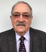
Prof Wolfgang Kiefer
Wolfgang Kiefer studied physics at Ludwig-Maximilians-University (LMU) Munich, Germany. After his Ph D in Physics he joined the Chemistry Division of the National Research Council in Ottawa, Canada, for postdoctoral work in the group of Dr Harold Bernstein and then returned to LMU Munich where he finished his habilitation in 1977. Shortly afterwards he became Professor of Physics at the newly founded University of Bayreuth, Germany, where he stayed until 1985. From 1985 to 1988 he was Full Professor for Physics and Head of Institute for Experimental Physics at Karl-Franzens-University of Graz, Austria. He finally accepted a chair in Physical Chemistry at University of Würzburg in 1988, where he stayed until his retirement in 2006. He also had been Vice-Dean and Dean of the Faculty for Chemistry and Pharmacy.
He was and still is member of several scientific journals (e.g. Applied Spectroscopy, Journal of Raman Spectroscopy (JRS), Chemical Physics Letters, Chinese Journal of Light Scattering, Spectroscopy Letters, Asian Journal of Physics, Asian Chemistry Letters), and he was Editor-in-Chief of JRS from 2000 to 2009. His research interests include laser physics, femtosecond coherent four-wave-mixing spectroscopy (e.g. Coherent Anti-Stokes Raman spectroscopy, CARS), studies of ultrafast ground and excited state dynamics in gases and in dense materials, coherent control of vibrations, nanosecond CARS, resonance Raman spectroscopy, development of theories for resonance Raman scattering, FT-IR spectroscopy and density functional theory calculations on metal complexes, Raman spectroscopy on non-crystalline samples (glasses, etc.), nanostructures, II-VI semiconductor quantum dots and quantum wires, biological and biochemical samples, polymers, and phase transitions, Raman-Mie scattering from micro-particles, surface enhanced Raman scattering (SERS), development of special techniques for Raman spectroscopy, matrix isolation spectroscopy, micro-Raman imaging and mapping, CARS microscopy. He has published more than 850 papers, among them more than 630 peer reviewed articles, 49 book articles, and he is co-author of 5 books. Out of his former research groups there had been 85 Ph D students from which 12 are now university professors in Germany, Romania, China, and USA.
He was visiting and is honorary professor of several universities and he received an honorary doctoral degree from Babes-Bolyai-University Cluj-Napoca, Romania. He also received several international awards, among them the prestigious Pittsburgh Spectroscopy Award and the first Raman Lifetime Award provided by the International Conferences on Raman Spectroscopy (IOCORS). During the celebration of 90 years of Raman effect on 28 February 2018, he had been honoured by the Indian Institute of Science in Bangalore for his “Lifetime Contribution to Raman Spectroscopy”. Other awards and honours are: Member of the Advisory Board of the Committee on Light Scattering of the Chinese Physical Society, Foreign Councilor of the Institute for Molecular Science, Okazaki, Japan, Honorary Fellow of the Laser and Spectroscopy Society of India, Distinguished Service Award of the Society for Applied Spectroscopy, Fellow of the Society for Applied Spectroscopy, USA. Several international journals have published special issues in honour of him (Asian Chemistry Letters, 2004, Asian Journal of Physics, 2006, 2016, and 2021, Journal of Raman Spectroscopy, 2006 and 2016, Zeitschrift für Physikalische Chemie, 2011). The Commemorative Issue of JRS (Vol. 37, No. 1-3, 2006) on the occasion of his 65th birthday contain a detailed profile of him and most of the titles of his publications. He has organized a great number of special issues for various international journals in the field of spectroscopy. He also had been chairman of the XIIIth ICORS, which took place in 1992 at University of Würzburg, Germany. Remarkably, he is the only person who attended all hitherto 26 ICORS meetings since the first one in 1969 in Ottawa, Canada. He is also Honorary Chairman of series of ICOPVS conferences founded by Prof Vinod Rastogi.
Since his retirement he runs a well-equipped private Raman laboratory in the basement of his house named Eisingen Laboratory for Applied Raman Spectroscopy (ELARS), now performing Raman spectroscopy as a hobby but still generating credible scientific output related to technological development.
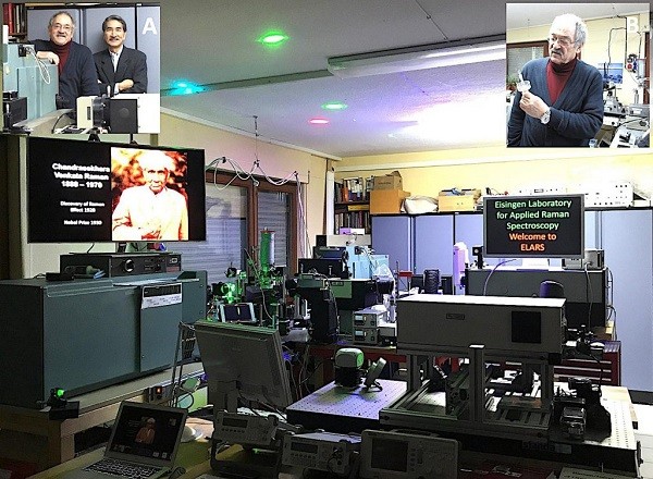
Inserts: A, left above: Professor Hiro-o Hamaguchi visiting ELARS in November 2013; B, right, above: Prof. Kiefer (2013), holding the original rotating Raman cell in his hands, which he developed at NRCC, Ottawa, in 1970; photo from H. Hamaguchi.
Message